Introduction to Zirconia Bridge Look
Zirconia bridges are a type of dental restoration that replaces missing teeth with an aesthetically pleasing and functional artificial solution. Crafted from zirconium dioxide, a high-strength ceramic material, these bridges are designed to mimic the appearance of natural teeth closely. Due to their impressive durability and resistance to wear, zirconia bridges are increasingly favored by dental professionals for long-term tooth restoration.
The primary purpose of a zirconia bridge is to restore both the functionality and appearance of the natural dentition. Whether used for single-tooth replacement or as part of a multi-unit restoration, these bridges serve to improve chewing efficiency, maintain the alignment of remaining teeth, and prevent bone loss in the jaw. They effectively bridge the gap created by one or more missing teeth, thereby enhancing the overall oral health of patients.
One of the significant advantages of zirconia bridges is their aesthetic appeal. The material’s translucency allows for a natural-looking color match with existing teeth, making these restorations less detectable. Additionally, zirconia bridges are known for their biocompatibility, which minimizes the risk of allergic reactions and ensures acceptance by the body. As a result, patients often prefer zirconia bridges over traditional metal-based options, which may not only be less appealing visually but also more prone to causing sensitivity.
In conclusion, the integration of zirconia bridges in dental restoration offers a reliable, functional, and visually appealing solution for patients with missing teeth. Their growing popularity reflects advancements in dental technology, which prioritize patient comfort, aesthetic quality, and long-lasting results.
Basics of Dental X-Rays
Dental X-rays are a crucial tool in modern dentistry, allowing practitioners to visualize the structure of the teeth, bones, and surrounding tissues beneath the surface. These images are essential for diagnosing a range of dental conditions, from cavities to periodontal diseases and even bone abnormalities. X-rays operate using a small dose of radiation to create images of the dental anatomy, effectively revealing details that cannot be seen during a standard oral examination.
There are several types of dental X-ray imaging techniques, each serving specific diagnostic purposes. The most common types include periapical, bitewing, and panoramic X-rays. Periapical X-rays capture the entire tooth structure, from the crown to the root, and are particularly useful for assessing dental abscesses or the health of bone around the teeth. Bitewing X-rays, on the other hand, show the crowns of the upper and lower teeth simultaneously, making them invaluable in detecting interproximal decay and monitoring the periodontal health of patients. Lastly, panoramic X-rays provide a broad view of the jaws, teeth, and surrounding structures, aiding in treatment planning for orthodontics or dental implants.
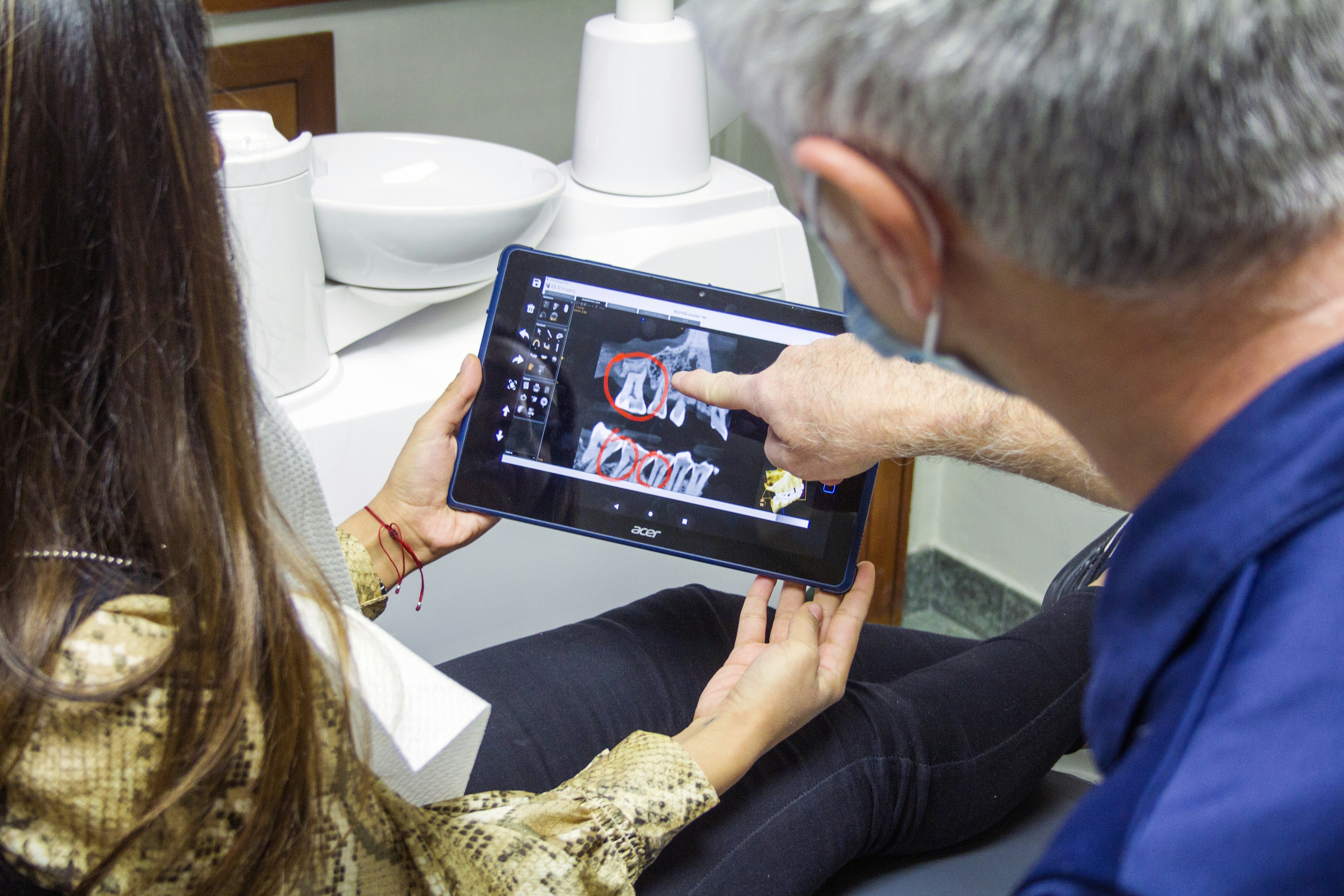
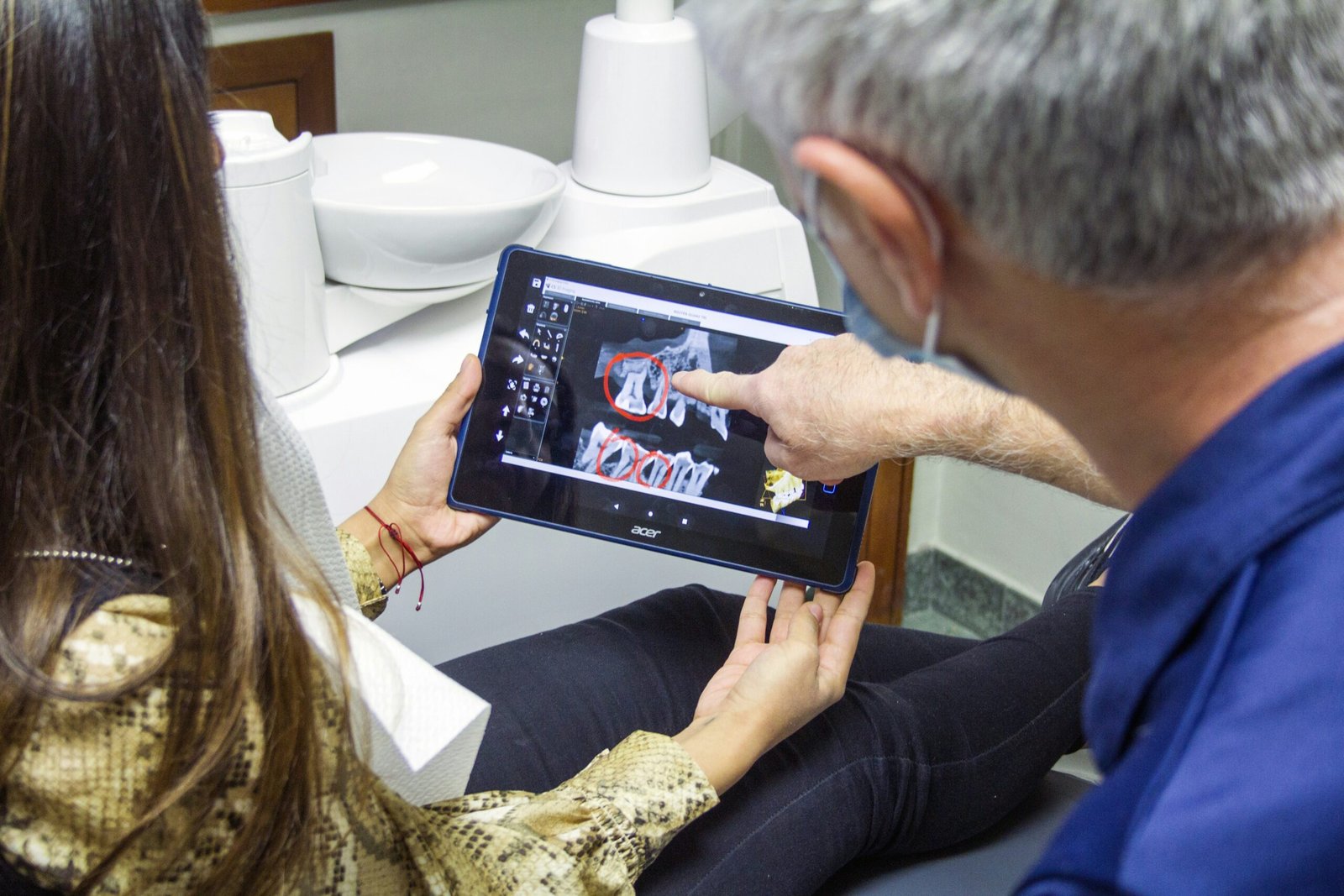
The significance of dental X-rays extends beyond merely identifying existing conditions; they also play a vital role in preventive care. Regular X-ray examinations can help detect issues before they manifest as more severe problems, ultimately preserving dental health and saving patients from extensive treatments. In addition, these images facilitate communication between dental professionals and their patients, allowing for a better understanding of the patient’s dental health status and the proposed treatment options.
Understanding Radiopacity
Radiopacity refers to the property of a material that determines how well it can obstruct the passage of X-rays. In dental imaging, radiopacity is crucial for identifying the density and composition of various structures within the oral cavity, including bones, teeth, and dental restorations. The varying levels of radiopacity mean some materials appear more opaque, while others are more transparent on an X-ray. This distinction is primarily due to the atomic number and density of the materials involved.
On an X-ray image, radiopaque materials appear white or light-colored because they absorb X-rays more effectively, preventing them from reaching the X-ray film or sensor. This characteristic is essential for diagnostic purposes, enabling dental practitioners to assess the integrity of the teeth and surrounding structures. For instance, metals like gold or silver are considered highly radiopaque due to their dense atomic structures. In contrast, softer tissues and air-filled spaces will appear dark because they allow X-rays to pass through with minimal absorption.
Zirconia, a popular material in dental restorations, such as bridges and crowns, has a notable radiopacity. While not as radiopaque as metals, zirconia exhibits sufficient density to be recognized on X-ray images, appearing as a distinct area in the dental arch. This enables dentists to monitor the condition of zirconia bridges over time, facilitating early detection of any potential issues related to the underlying tooth structure or periodontal health. Understanding how radiopacity affects the visibility of zirconia on X-rays is essential for both dental professionals and patients, as it reinforces the role of imaging in effective dental care.
The Appearance of Zirconia on X-Rays
Zirconia, a popular material in dental prosthetics, has distinct characteristics when observed through X-ray imaging. Unlike natural teeth, which exhibit varying shades of radiopacity due to their organic and inorganic components, zirconia appears as a dense, uniform structure on radiographs. This material generally presents with a high degree of radiopacity, meaning it absorbs X-ray radiation more effectively than surrounding bone and soft tissue.
On an X-ray, natural teeth, composed of enamel, dentin, and pulp, can show gradients of density, which is essential for their diagnostic evaluation. Enamel, being the hardest tissue, appears lighter, while dentin is slightly darker. In contrast, zirconia does not showcase these gradients. Instead, its consistent radiopacity can make it appear notably more pronounced against surrounding structures. This difference can sometimes complicate the interpretation of scans, as the clear demarcation between zirconia and alveolar bone might lead to confusions regarding the anatomy.
Furthermore, zirconia’s homogeneity can create artifacts on X-rays, which dental professionals need to be aware of during diagnosis and treatment planning. Since it does not exhibit any radiolucency, indicating the presence of decay or infection that can be commonly seen near natural teeth, understanding how zirconia shows up on radiographs is crucial for evaluating overall oral health. Dental professionals must therefore interpret X-rays with the knowledge of these distinct features to make accurate assessments regarding the surrounding structures and the integrity of the zirconia bridge itself.
Why Zirconia is Radiopaque
Zirconia, or zirconium dioxide (ZrO2), is recognized for its unique properties that contribute to its use in dental applications, particularly in creating durable and aesthetic restorations like zirconia bridges. One of the fundamental characteristics of zirconia is its radiopacity, which allows it to appear distinctly on dental X-rays. This quality is crucial for clinicians as it aids in diagnosing and monitoring the condition of dental structures.
The radiopaque nature of zirconia is primarily attributed to its high atomic number. Materials with higher atomic numbers tend to absorb more X-ray radiation, leading to a denser appearance on radiographic images. Zirconia’s atomic composition enables it to stand out against surrounding tissues and other dental materials that may be less radiopaque. This differential visibility helps dentists assess the integrity of zirconia bridges and ensure their proper placement within the dental arch.
Moreover, zirconia exhibits superior strength and aesthetic properties, making it a preferred choice over traditional materials such as metal or resin. When viewed on X-rays, zirconia’s radiopaque characteristics facilitate the identification of any marginal gaps or issues related to fit and adaptation that may not be immediately visible during clinical examinations. The clear distinction on X-ray images ensures that dentists can make informed decisions regarding treatment plans and necessary interventions.
In conclusion, the radiopaque nature of zirconia plays a pivotal role in dental imaging, enabling practitioners to effectively evaluate dental restorations. Its unique properties not only enhance visibility on X-rays but also contribute to the overall success of dental treatment outcomes.
Detecting Underlying Health Issues with X-Rays
Dental X-rays play a pivotal role in modern dentistry, especially in the context of evaluating the integrity of restorative work, such as zirconia bridges. These imaging techniques allow dentists to visualize not only the bridges but also the underlying structures of the teeth and jawbone, providing critical insights into a patient’s dental health that may not be visible during a routine examination.
When a zirconia bridge is placed over natural teeth or implants, some potential issues may be obscured from direct visual inspection. For instance, secondary caries, which may form at the margins of the bridge, can be challenging to detect without advanced imaging. X-rays help in identifying such decay by revealing any anomalies in the dental structure beneath the bridge. This is crucial for timely intervention, as untreated decay can lead to more severe complications, including tooth loss.
Moreover, X-rays are essential for identifying periapical infections or bone loss around the roots of supporting teeth or implants. Conditions such as periodontitis can affect the foundational support for a zirconia bridge, and early detection through X-ray imaging can aid in preventing further deterioration and maintaining oral health. Additionally, X-rays serve to assess bone density, ensuring that the jawbone is healthy enough to support any restorative work.
In conclusion, X-ray imaging is a vital tool in dental diagnostics, particularly for patients with zirconia bridges. By enabling dentists to look beyond the visible, X-rays help in identifying underlying health issues, facilitating prompt and effective treatment, and ultimately, ensuring the longevity of dental restorations and overall oral health.
Benefits of Zirconia Bridges in Dental Imaging
Zirconia bridges have emerged as a preferred choice for dental restorations due to their numerous advantages, particularly regarding dental imaging practices. When considering how a zirconia bridge looks on an X-ray, one must first acknowledge the material’s unique properties. Zirconia is known for its high biocompatibility and excellent mechanical strength, which makes it an ideal candidate for dental structures that require durability and aesthetic appeal.
One of the primary benefits of zirconia in dental imaging is its minimal interference with radiographic diagnosis. Unlike traditional metal-based restorations, which might obscure details on X-rays, zirconia offers clearer imagery due to its radiolucent properties. This means that when a dentist examines X-rays, a zirconia bridge will not hinder the visibility of surrounding dental structures, allowing for more accurate assessments of both hard and soft tissues. The reduced interference can help in early detection of potential issues such as decay beneath the bridge or periodontal problems, ultimately leading to improved patient outcomes.
Moreover, zirconia bridges present a comprehensive solution in aesthetic dentistry. They replicate the natural translucency of teeth far more effectively than metals, which can appear opaque on X-rays. This resemblance enhances both appearance and functionality, as patients can expect lifelike integration of the restorations with their existing dentition. Consequently, the aesthetic results are visually appealing, contributing to overall patient satisfaction and self-esteem.
In addition, the longevity and resistance to wear and tear that zirconia bridges offer further emphasize their value in the dental field. As materials employed in dental restorations continue to evolve, zirconia stands out as a premier option, combining functionality with superb imaging characteristics.
Limitations of X-Rays in Assessing Zirconia Bridges
X-rays are a fundamental tool in dental imaging, providing crucial insights into the condition and placement of dental structures, including zirconia bridges. However, there are several limitations associated with the use of X-rays for evaluating these bridges, which can impact the diagnostic accuracy and clinical decision-making process.
One major limitation is that zirconia, known for its strength and biocompatibility, is less radiopaque than metal materials traditionally used in dental restorations. This property can lead to challenges in identifying the precise outline and integrity of a zirconia bridge on X-ray images. Consequently, ambiguous or indistinct images may result in potential misinterpretations regarding the condition of the bridge and the surrounding dental structures.
Additionally, X-rays predominantly reveal a two-dimensional view of three-dimensional structures. This limitation may obscure important details, such as the interfaces between the zirconia bridge and natural teeth or implants, leading to misdiagnosis of issues like marginal bone loss or other complications. A proper assessment may require alternative imaging techniques that provide multi-dimensional perspectives, such as cone beam computed tomography (CBCT), which can deliver greater clarity in understanding the exact position and condition of the zirconia bridge.
Furthermore, the presence of dental artifacts, which can arise from materials used in restorations or prior treatments, might also distort the diagnostic-quality of the X-ray images. Such artifacts can complicate the interpretation of the images, making it difficult for dental professionals to discern between normal anatomical variations and actual pathological changes.
In light of these limitations, it is essential for dental practitioners to recognize when additional imaging techniques are warranted. This awareness ensures a comprehensive approach to evaluate zirconia bridges, ultimately enhancing patient care and treatment outcomes.
Conclusion and Future of Zirconia in Dentistry
In conclusion, zirconia bridges have demonstrated a significant transformation in dental practices, particularly in how dental imaging captures their presence during diagnosis and treatment planning. These bridges, recognized for their aesthetic appeal and biocompatibility, have gained acceptance, revolutionizing restorative dentistry. The ability of zirconia to blend seamlessly with natural dentition ensures not only functional restoration but also a pleasing appearance, which is essential for patient satisfaction.
Through our exploration of dental imaging, it is evident that systems such as X-rays play a fundamental role in evaluating the integrity and positioning of zirconia bridges. X-ray imaging provides essential insights into the structural alignment and integrity of these restorations, making it an indispensable tool for dental professionals. While zirconia is radiopaque, allowing for clear visualization on X-rays, the future may bring innovations in imaging technologies that enhance detail and clarity, propelling forward the standard of care.
Looking ahead, advancements in material sciences are poised to further improve the properties of zirconia. The potential introduction of new zirconia formulations may enhance their strength and translucency, promoting even greater durability and aesthetic outcomes. Additionally, research into refined imaging techniques may render even clearer images of zirconia restorations, aiding practitioners in precise evaluations.
Furthermore, the ongoing development in digital dentistry—including computer-aided design (CAD) and computer-aided manufacturing (CAM)—may streamline the creation of zirconia restorations while ensuring accurate fits and finishing. As technology continues to evolve, the role of zirconia in restorative procedures will likely become increasingly significant, setting new benchmarks in dental excellence.